© 2012 Matteo Lencioni | P.I. 00897520466

eng

GENERAL INFORMATION ON MELANOMA
Melanoma is a malignant neoplasm which is most frequently found on the skin. The incidence and death rate of this type of tumour is continuously increasing throughout the world.
Melanoma is diagnosed clinically. The pigmented lesions on the skin are tested using a dermoscope and the development of a melanoma is avoided. (so-called mole mapping)
Melanoma tumours which are not diagnosed and treated early on may be difficult to recover from.
Every year in Italy around 35000 people develop a melanoma and around 7300 die as a result. Over the past 10 years, the cases of melanoma diagnosed have doubled.
If diagnosed in time the melanoma can be treated. A melanoma is a tumour which results from melanocytes (the skin cells that contain the pigment called melanin). Melanoma is formed of melanocytes which transform into tumour cells following mutations provoked by external agents (UV and ionising radiation) which are more frequent in individuals who are genetically predisposed (family history of melanoma). If not treated in time, melanoma has a high possibility of spreading.
The lesion may appear unexpectedly on healthy skin or may originate from a preexisting nevus, which may be congenital. The areas which are most at risk are generally those which are most exposed (the torso for males and the legs and arms for females). The most known risk factor is sun exposure, above all in those with light skin or red and blonde hair and blue or green eyes (the so-called phototypes at risk). Some large congential nevi may have a higher risk of developing into a melanoma.
Other risk factors are:
1) having a large number of nevi (more than 50)
2) having repeated, serious sunburns in childhood and adolescence
3) having one or more cases of melanoma in the family
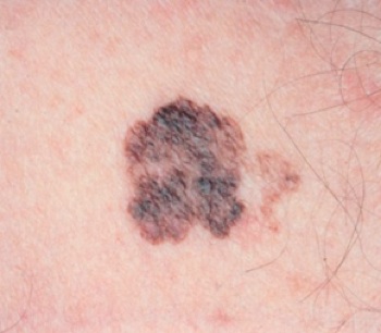
There are some rules to help distinguish a melanoma from a normal mole:
The ABCDE is a simple set of rules which the patient may follow alone to evaluate one's own moles as a first step. This involves paying attention to the following characteristics of the nevus, which if present (above all, more than two or three) requires the individual to report to their GP who will decide whether to refer them to a specialist.
A (asymmetry) - the lesion is not regular, it is not round but asymmetric and its two halves are not symmetrical
B (border) - the edge of the lesion is not regular but blurry
C (colour) - the colour is not uniform, but there are darker, irregular areas, with blackish, bluish, greyish or reddish tones, alternating with lighter areas (mottled appearance)
D (diameter) - usually an abnormal nevus is 0.5 mm or larger
E (evolution) - very important; if a nevus changes aspect and/or grows, in the space of a few days an evaluation is required. This does not mean that all nevi which grow are melanoma, but melanoma normally changes suddenly and changes the preexisting mole.
Melanoma


Specialist in Plastic Reconstructive Aesthetic
